
The human eye is the structure around which the entire optical industry revolves. There are many reasons why such a large industry would develop around this seemingly simple part of human anatomy. First, much of what we understand about the world around us has been developed over the course of many years through our sense of sight. While the brain is the structure that processes, interprets, and responds to the environment, it relies heavily on the signals it receives from other parts of the body.
Our eyes start sending signals to our brain from the moment we enter the world. These signals allow the brain to develop in such a way that gives us the ability to move about our environment without crashing into things or letting external forces harm us. The importance of this process has been demonstrated in cases where sight has been restored to individuals who have been blind from a very young age. Once vision has been restored in these people, their brain must learn things that most people take for granted such as depth perception and object relativity. This process can take some time because the brain was not able to develop like it normally would have.
While most of us do not consciously consider the importance of our vision on a regular basis, we are quick to realize how important it is to us once it has become severely impaired or eliminated. In fact, vision loss is one of the most difficult medical problems for people to accept. This is because, along with the loss of sight, we lose the ability to perform many of the tasks that we depend on throughout our lives including driving, nonverbal communication, and visual inspection. The inability to live life, as we once understood it, is extremely depressing and can have a negative impact on our mental health and general well-being.
If we consider our vision, in the context of our daily lives, then it becomes profoundly obvious how a multi-billion dollar industry could have evolved to serve the medical and aesthetic needs associated with the eyes. The human eye is a truly unique aspect of our human anatomy that has given rise to an industry unlike any other in the healthcare community. The combination of physiological dependence and fashion significance has created a tremendous amount of opportunity for individuals who are well-trained to understand the eyecare needs that exist and who can provide high-quality products and services.
In order to develop a better understanding of the visual needs that people have, it is important to have an appreciation for the anatomy of the human eye. The following is a brief outline of the most common structures that opticians should know in order to pass the Certification Exams and to provide the highest possible level of care.
Anatomy of the Human Eye
The Three Layers:
- Fibrous Tunic (tunica fibrosa oculi)
- Sclera – the membrane responsible for maintaining the shape of the bulb.
- Cornea – the transparent dome that projects in front of the sclera.
- Vascular Tunic (tunica vasculosa oculi)
- Choroid – this is a thin membrane consisting of small arteries and veins that serve to provide nutrition to the retina and convey vessels and nerves to the ciliary body and iris.
- Ciliary Body – consists of the orbiculus ciliaris, ciliary processes, and ciliaris muscle. The ciliaris muscle is the main agent in accommodation.
- Iris – this is a thin contractile disk that is suspended in the aqueous humor in between the cornea and the lens. It is perforated by the pupil and distinguishes the anterior chamber from the posterior chamber.
- Nervous Tunic (tunic interna)
- Retina – this is a nervous membrane where images are received. This is what people are referring to when they talk about the rods and cones.
The Three Chambers:
- Anterior Chamber – this is the fluid filled space between the iris and the cornea.
- Posterior Chamber – this is the space between the iris and the lens.
- Aqueous Humor – this is the gelatinous fluid that is secreted into the anterior and posterior chambers between the lens and the cornea. This should not be confused with the vitreous humor which exists in the larger space behind the lens. The aqueous humor has many functions including maintaining intraocular pressure and providing nutrients.
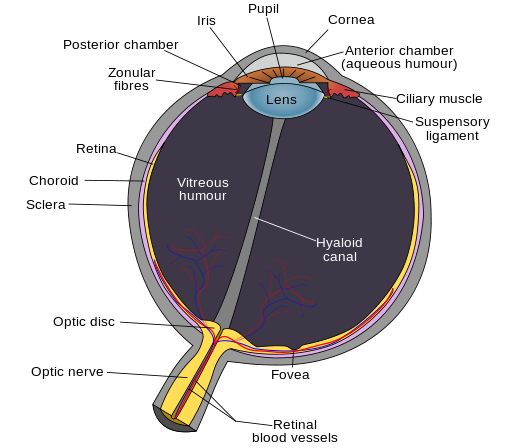
Lens – this structure is important in focusing light waves to create clear images. This process is commonly known as refraction and is associated with errors such as myopia, hyperopia, astigmatism, and presbyopia.
Canal of Schlemm – this structure is a tube that collects aqueous humor from the anterior chamber and transports it to the blood via the anterior ciliary veins. This structure is of significant importance because it can obstruct outflow of aqueous humor resulting in increased intraocular pressure which is common in Glaucoma.
Vitreous Humor – this is the gelatinous fluid that fills the space between the lens and the retina. Floaters result from blood, cells, or other debris that has managed to get into the vitreous humor. This gel also aids in maintaining the shape of the eye.
Optic Nerve – this is cranial nerve two which is responsible for transmitting visual information from the retina to the brain. The ‘Blind Spot’ phenomenon results from the absence of photo receptors in the area where the optic nerve exits the eye.
The Six Extraocular Muscles:
- Medial Rectus – toward the nose
- Lateral Rectus – away from the nose
- Superior Rectus – upward, toward the nose, inward
- Inferior Rectus – downward, away from the nose, inward
- Superior Oblique – toward the nose, downward, outward
- Inferior Oblique – away from the nose, upward, outward
Click Here to read about light and the visual pathway.